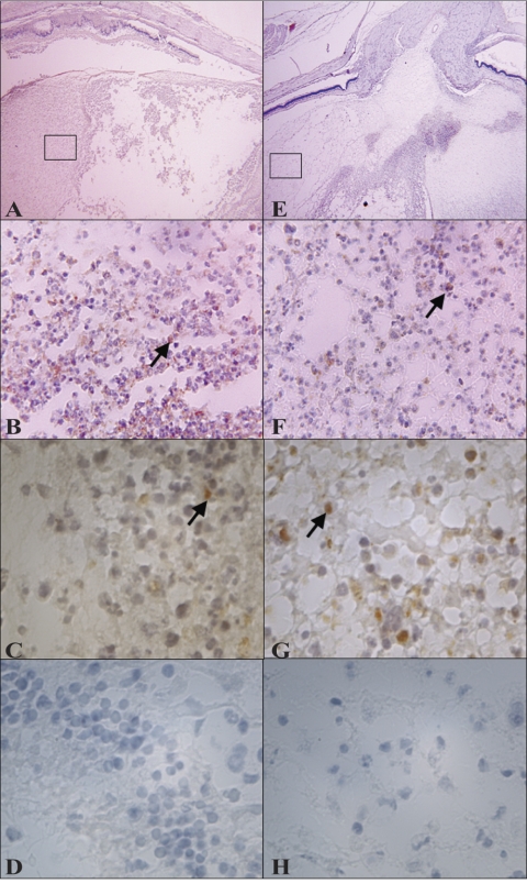
Figure 6.
Representative histology pictures (macrophage staining) of eyes infected intravitreously with the parent strain (A–D) or the mutant strain (E–H) at 48 hours PI. Arrows indicate macrophages (stained brown). (C, G) Magnifications of (B) and (F), respectively, to show morphology. (D, H) Isotype controls. Original magnification, 20× (A, E); 400× (B, F); 1000× (C, D, G, H). Areas from (A) and (E) magnified in (B) and (F) are boxed.

