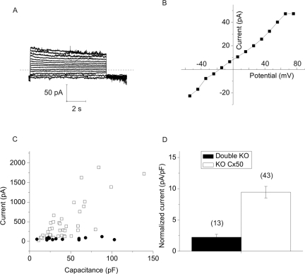
Figure 9.
Loss of Cx50 and Cx46 abolishes calcium-sensitive membrane currents in single fiber cells. (A) Example of membrane currents recorded from a double KO fiber in divalent cation-free NaGluconate Ringer's solution. The voltage clamp protocol consisted of sequential steps from a holding potential of −60 mV to 80 mV in 10 mV increments. Dashed line: zero current. (B) I–V relations obtained from the data shown in (A). The current was measured at the end of the 8-second pulse and plotted as a function of voltage. (C) Steady state current (measured at the end of an 8-second pulse to 80 mV) plotted as a function of membrane capacitance for the double KO cells (solid circles) and the KOCx50 cells (open squares). (D) Histogram compares the mean area-specific current of the double KO cells (open bar) and the KOCx50 cells (black bar). The area-specific current was calculated from the data shown in (C), assuming an area-specific membrane capacitance of 1 μF/cm2. Number of cells is indicated in parentheses.

