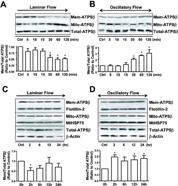
Figure 1. Flow induces ATPSβ translocation between plasma membrane and mitochondria in ECs.

(A,B) Confluent monolayers of BAECs were subjected to laminar flow (12 dyne/cm2) or oscillatory flow (0.5±4 dyne/cm2) up to 2 hr, or kept as static controls. (C,D) The experimental conditions were the same as in A,B except the flow duration was up to 24 hr. Cells were lysed into 3 fractions (membrane, mitochondria and whole-cell lysates), and then ATPSβ was detected by western blotting. Flotillin-2 and MtHSP75 were also detected as markers for membrane or mitochondria fractions, respectively. Graph shows the ratio of membrane ATPSβ to total ATPSβ. The ratios of the static control were set to 1. Data were from 3 independent experiments. *P<0.05.
