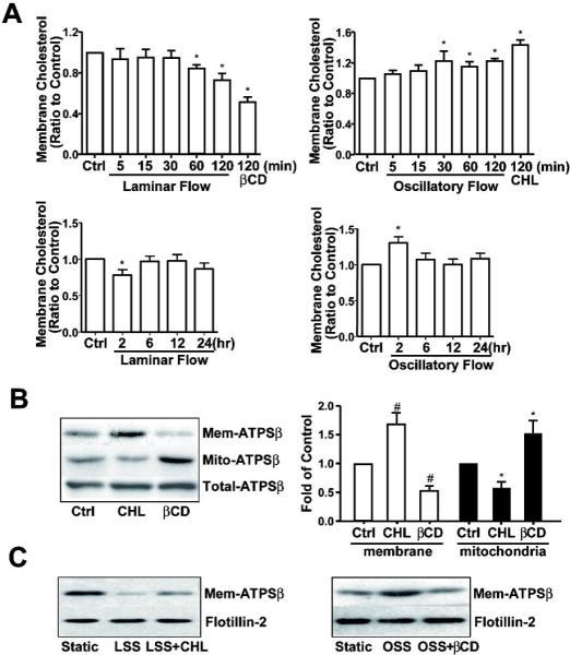
Figure 2. Endothelial membrane cholesterol plays an important role in flow-induced ATPSβ translocation.

(A) BAECs were subjected to laminar flow or oscillatory flow for different times. βCD (5 mM, 2 hr) or cholesterol (CHL, 30 μg/ml, 2 hr) was applied as positive controls. The level of membrane cholesterol was determined with the same amount of protein, which was then normalized to that in the static control, set as 1. Data represent the mean±SEM from 3 independent experiments. * P<0.05 versus control; (B) BAECs were stimulated with CHL (30 μg/ml, 2 hr) or βCD (5 mM, 2 hr). Proteins from membrane or mitochondrial fractions and whole-cell lysates were isolated, and ATPSβ was detected by western blot analysis. Graph shows the ratio of membrane or mitochondrial ATPSβ to total ATPSβ, with that of static controls set as 1. The data are averaged from 3 independent experiments. # P<0.05 versus membrane control; *P<0.05 versus mitochondrion control. (C) In the presence of cholesterol or βCD, BAECs were subjected to laminar flow or oscillatory flow. Proteins from membrane fraction were isolated, and ATPSβ was detected by western blot analysis. Data were representative from 3 independent experiments.
