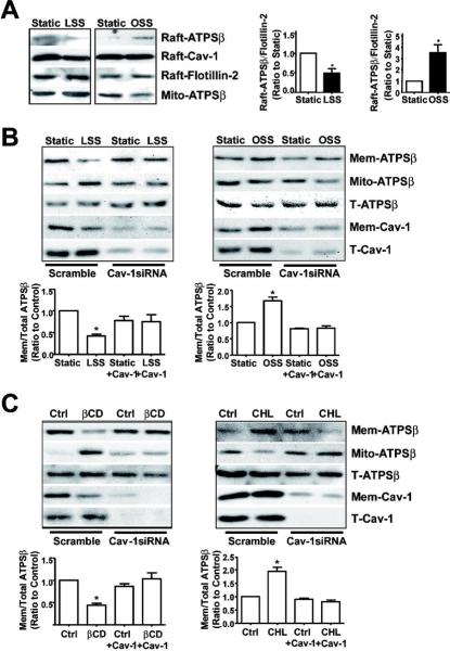
Figure 3. ATPSβ translocation depends on caveolin-1.
(A) BAECs were subjected to laminar flow or oscillatory flow for 2 hr. Sucrose gradient ultracentrifugation was used for isolating lipid rafts (fractions 4–5) and mitochondrial fractions (fractions 8–10). Lipid raft and mitochondrial fractions were examined by western blot analysis with anti-ATPSβ, anti-Cav-1, and anti-Flotillin-2 antibodies. The images are representative of 3 independent experiments. (B,C) BAECs were transfected with scramble or Cav-1 siRNA for 48 hr, then subjected to different flow patterns, βCD, or CHL for 2 hr. The level of ATPSβ and Cav-1 was detected by western blot analysis. Graph shows the ratio of membrane ATPSβ to total ATPSβ. Data were from 3 independent experiments with static control set as 1. *P<0.05. T: total; Raft: lipid raft; Mem: membrane; Mito: mitochondria.

