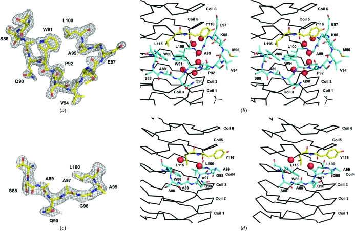
Figure 2.
Structure of the AlbG β-helix loop excursion. (a) Shaken F o − F c OMIT map (2.5σ) for the region bounding the loop adjoining coils 4 and 5 on face 4. (b) Stereo diagram of the loops position relative to the β-helix. Residues from the loop (86–100) are shown as sticks with cyan C atoms, whilst structural residues (Leu115 and Tyr116) from coil 5 are shown as sticks with yellow C atoms. Waters are shown as red spheres. (c) Shaken F o − F c OMIT map (2.5σ) for corresponding residues after reconstruction of the sequence to remove the β-helix loop excursion (AlbGΔ91–97). (d) Stereo diagram of the AlbGΔ91–97 structure about the deletion. For the OMIT maps, residues 87–100 were removed from the structure and the coordinates were randomly shaken by 0.3 Å, which was followed by a round of steepest-gradient refinement in PHENIX.

