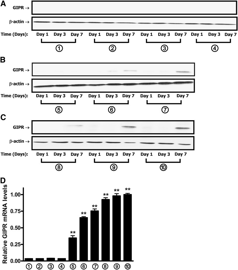
Fig. 2.
GIPR expression increases during adipocyte differentiation in 3T3-L1 cells. Protocols for treatment of 3T3-L1 preadipocytes with GIP are as described in the legend to Fig. 1A. A–C: The time course of GIPR protein expression during 3T3-L1 adipocyte differentiation is shown. Western blot analyses were performed using antibodies against GIPR and β-actin. D: GIPR mRNA expression during 3T3-L1 adipocyte differentiation is shown. 3T3-L1 preadipocytes were treated for 7 days, as described in the legend to Fig. 1A, and real-time RT-PCR was performed with extracts to quantify GIPR mRNA levels, shown as fold difference versus control normalized to 18S rRNA expression levels. All data represent three independent experiments, each carried out in duplicate, and Western blots are representative of n = 3 replicates. Significance was tested using ANOVA with Newman-Keuls post hoc test, where ** represents P <0.05 versus control group, treated with conditional culture medium 1 (no IBMX/Dex/insulin/GIP additions) for 7 days.

