Figure 1.
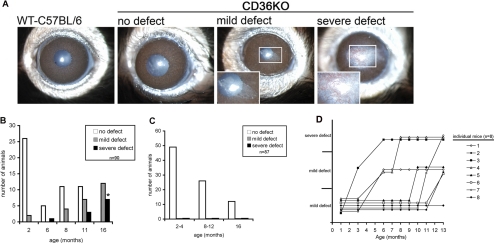
CD36−/− mice spontaneously developed corneal defects that increase in frequency and severity with age. (A) Slit lamp pictures of representative eyes from of CD36−/− mice that display (1) no corneal defect, clear cornea; (2) mild corneal defect, mild corneal haze through which the iris is visible; and (3) severe corneal defect, corneal opacity, and neovascularization through which the iris is not visible. (B) The frequency of corneal defects in CD36−/− mice (n = 90). (C) Age-matched WT C57BL/6 mice served as a negative control (n = 87). (D) Disease progression was monitored monthly in a subset of CD36−/− mice (n = 8). Asterisk indicates significant difference in the frequency of severe lesions between 16-month-old CD36−/− and WT-C57BL/6 mice (P = 1.745 × 10⁻⁹) and was determined by Fisher's exact test. Development of corneal defects in CD36−/− mice was age dependent as determined by proportional odds logistic regression (beta = −0.3652, P = 5.005 × 10⁻⁸).

