Figure 2.
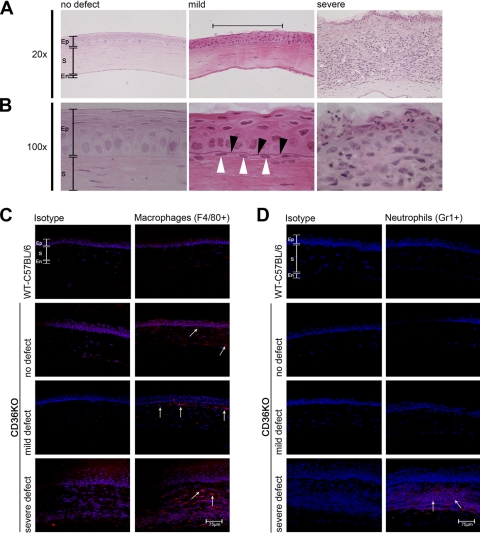
Histology of corneal defects. Corneas were recovered from mice with no defects, young CD36−/− mice; mild defects; or severe defects. (A, B) Corneas were fixed, paraffin embedded, sectioned, and stained with hematoxylin and eosin. (C) Corneas were recovered from WT C57BL/6 mice or CD36−/− mice with either no defect, mild defect, or severe defects. Corneas were snap frozen in OCT, sectioned, and stained with either an isotype-matched control antibody or an anti-F4/80 antibody (specific for macrophages). (D) A similar set of corneal sections were stained with either an isotype-matched control antibody or an anti-GR1 antibody (specific for neutrophils). All sections were stained with a cyanine nucleic acid stain (blue) to identify the cell nucleus. Immunofluorescence was examined via confocal microscopy. Ep, epithelium; S, stroma; En, endothelium. Black arrowheads identify a thickened basement membrane. White arrowheads identify a stromal infiltrate directly under the mild corneal defect. White arrows identify F4/80- and GR1-positive cells.

