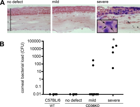
Figure 3.
Detecting bacteria in corneal defects. Corneas were recovered from WT-C57BL/6 mice or CD36−/− mice with either no defects, mild defects, or severe defects. (A) Corneal sections were stained with a Gram stain. Gram-positive bacteria (dark purple) were detected in corneas with severe lesions. Pictures are representative of corneas from at least three different mice. (B) The bacterial load within individual corneas was determined by homogenizing and culturing the corneal tissue in BHI agar cultures. Bacterial colonies were enumerated and the CFUs for each cornea displayed [n = 4 (C57BL/6), n =4 (no defect), n = 8 (mild defect), n = 4 (severe defect)]. Asterisk indicates significant increase in bacterial load compared with all other groups (Fisher's exact test, P = 0.01). Ep, epithelium; S, stroma; En, endothelium. Black arrows identify Gram-positive cocci.

