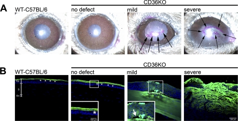
Figure 4.
The epithelial barrier function in the corneas of CD36−/− mice. (A) Rose Bengal staining was performed on WT-C57BL/6 mice and CD36−/− mice with different defects. Rose Bengal stains positive (pink) for disrupted mucin layers. (B) Epithelial tight junctions were examined with LC-biotin. Intact epithelial tight junctions prevent LC-biotin from penetrating the cornea and staining the stroma (WT-C567BL/6 and CD36−/− mice with no defect). Disrupted epithelial tight junctions allow LC-biotin to penetrate and stain the stroma (CD36−/− mice with mild and severe defects). All sections were counterstained with a cyanine nucleic acid stain (blue nuclei) and analyzed via confocal microscopy. Pictures are representative of five mice per group. Ep, epithelium; S, stroma; En, endothelium. Black arrows identify the positive Rose Bengal staining in CD36−/− corneas with mild and severe defects. Asterisks identify the LC-biotin (green) sitting on top of an intact epithelial barrier in WT-C57BL/6 and young CD36−/− mice with no defect. White arrow identifies epithelial detachment from the basement membrane.

