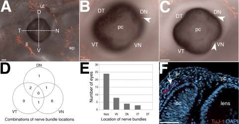
Figure 1.
Innervation of the anterior ocular region at E12.5. (A) An embryo showing innervation of the upper and lower eyelids, and the whisker pad but no innervation of the eye. To determine the positioning of nerve bundles before cornea innervation, the eye was divided into quadrants along the dorsal-ventral and temporal-nasal axes. (B) Innervation of the DN quadrant (arrowhead). (C) Innervation of the DN and VN quadrants (arrowheads). (D) Venn diagram summarizing the number and overlap of innervated quadrants. (E) Quantification of eye innervation and position of pioneer nerve bundles. (F) Cross-section through an E12.5 eye counterstained with DAPI showing a nerve bundle (arrow) in the periocular region projecting toward the presumptive cornea. Figure 1F was imaged from a similar location as the boxed region in Figure 2D. D, dorsal; N, nasal; V, ventral; T, temporal; DN, dorsal-nasal; VN, ventral-nasal; ll, lower eyelid; ul, upper eyelid; wp, whisker pad; pc, presumptive cornea; pr, periocular region; oc, optic cup. Scale bars: (A–C) 100 μm; (F) 50 μm.

