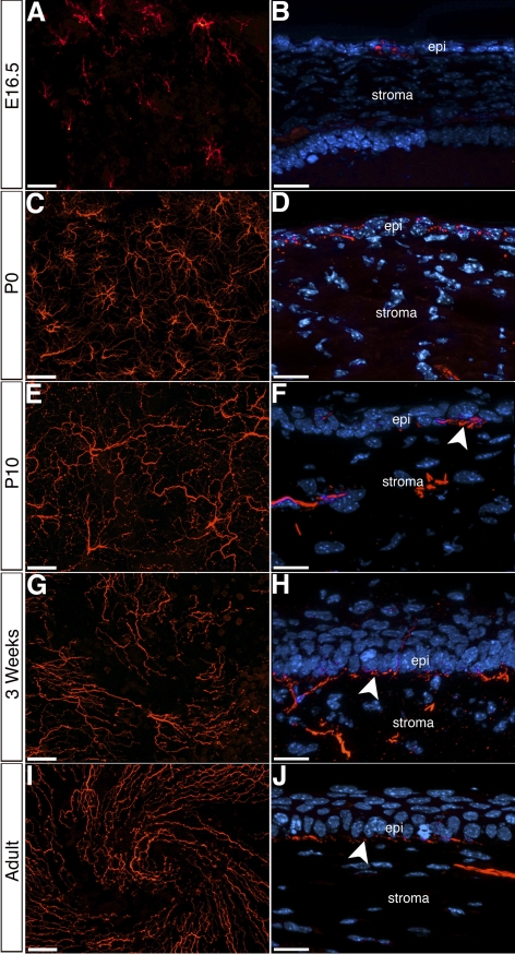
Figure 4.
Innervation of the mouse corneal epithelium. (A, C, E, G, I) Whole-mount immunostaining of cornea epithelia showing the distribution of epithelial nerves at the corneal apex during different stages of development and in adult. (B, D, F, H, J) Cross-sections of corneas at corresponding time points, showing innervation of the epithelium and the formation of the subbasal plexus (arrowheads). epi, epithelium; st, stroma. Scale bars: (A, C, E, G, I) 50 μm; (B, D, F, H, J) 20 μm.

