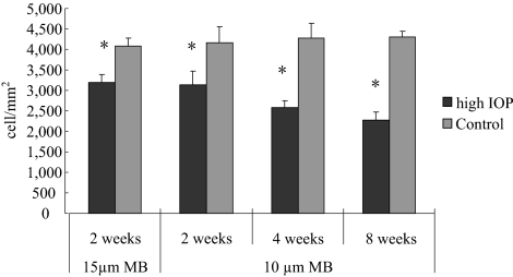
Figure 7.
Quantification of RGC loss after IOP elevation. RGC counts in retinal sections of mice that received microbead injection, in which RGCs were labeled with anti–β-III-tubulin immunohistochemistry. Mice that received anterior chamber injection of 15 μm microbeads were killed at 2 weeks (n = 10), and those that received 10 μm microbeads were killed at 2 (n = 8), 4 (n = 6), and 8 (n = 8) weeks after injection. Mice killed at week 8 received a second injection of microbeads at week 4 to maintain the elevated IOP. RGC counts taken from the uninjected contralateral eyes were used as controls. *P < 0.001 compared with the corresponding controls by Student's t-test.

