Figure 1.
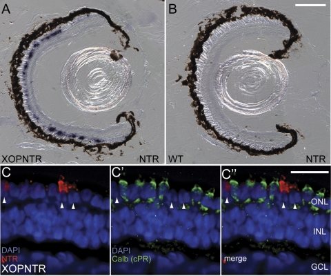
NTR expression in rod photoreceptors of stage 50 F1 XOPNTR tadpoles. (A, B) In situ hybridization was used to detect NTR expression (purple) in transgenic and control tadpoles. NTR expression was detected in the outer nuclear layer of XOPNTR tadpoles (A) but was undetectable in wild-type sibling embryos (B). (C–C″) Retinas were triple stained to label NTR-expressing cells (Fast red; red), cones (calbindin; green), and nuclei (DAPI; blue). NTR-expressing cells (C, arrowheads) do not express calbindin (C′, C″). Scale bars: 100 μm (A, B); 25 μm (C–C″).

