Figure 4.
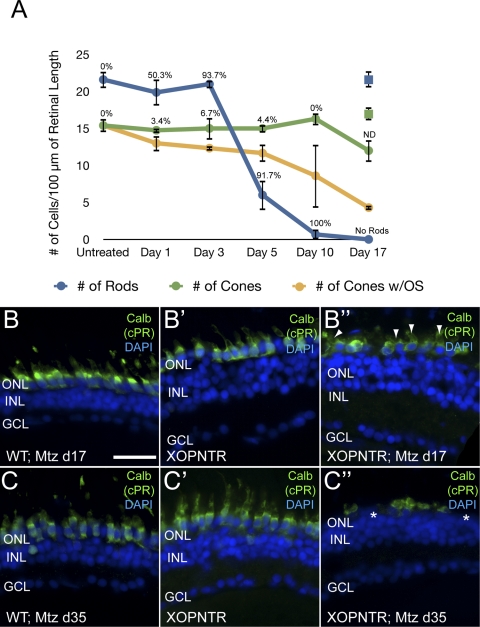
Cones degenerate after rod ablation. Stage 52 XOPNTR tadpoles were treated with Mtz for 1 to 17 days. Retinal sections were stained for rods (transducin), cones (calbindin), apoptotic cells (TUNEL), and nuclei (DAPI). The blue, green, and orange lines indicate the number of rods, cones, and cones with outer segments over time, respectively. The percentage of TUNEL-labeled cells at each time point is shown. The numbers of rods (blue) and cones (green) in control wild-type animals treated with Mtz for 17 days are shown for comparison (all cones had outer segments). Similar results were observed in transgenic control animals treated with DMSO for 17 days (18.3 ± 0.9 rods; 16.3 ± 0.9 cones). Retinal sections of wild-type Mtz-treated (B, C), XOPNTR untreated (B′, C′) and XOPNTR Mtz-treated (B″, C″) tadpoles were stained for calbindin. Animals were treated for either 17 (B, B″) or 35 (C, C″) days. All sections were counterstained for nuclei (DAPI). Asterisks: region lacking an outer plexiform layer (C″). Arrowheads: cones with outer segments. ND, not determined. Scale bar, 20 μm.

