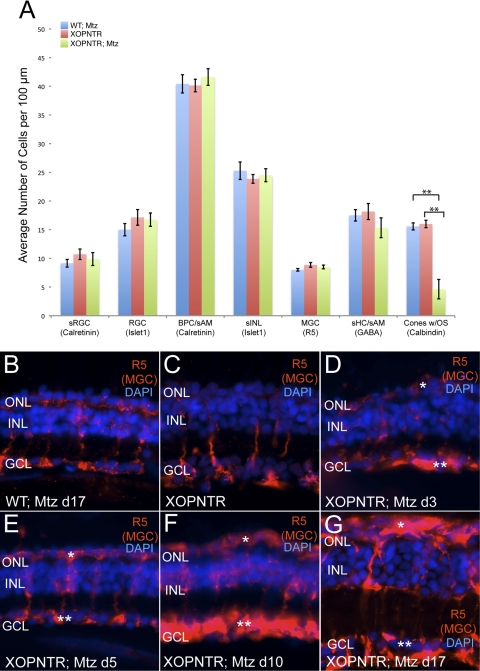
Figure 5.
Secondary changes follow rod photoreceptor ablation. The retinas of control and XOPNTR stage 52 tadpoles treated with Mtz for 17 days were immunostained with retinal cell markers. (A) Blue, red, and green bars indicate the average number of cells detected in wild-type Mtz-treated, XOPNTR untreated, and XOPNTR Mtz-treated tadpoles for each respective marker. (B–G) Retinal sections of wild-type Mtz-treated (B), XOPNTR untreated (C), and XOPNTR Mtz-treated (D–G) tadpoles stained for R5 (Müller glia). Animals were treated for 3 (D), 5 (E), 10 (F), and 17 (G) days. All sections were counterstained for nuclei (DAPI). Single asterisks: Müller cell gliosis spreading to the subretinal layer. Double asterisks: Müller cell gliosis spreading to the GCL. RGC, retinal ganglion cells; sRGC, subset of retinal ganglion cells; BPC, bipolar cells; sAM, subset of amacrine cells; sHC, subset of horizontal cells; sINL, subset of inner nuclear layer cells; MGC, Müller glial cells. Mean ± SEM is indicated. **P < 0.0001. Scale bar, 20 μm. See also Supplementary Fig. S1, http://www.iovs.org/lookup/suppl/doi:10.1167/iovs.10-5347/-/DCSupplemental.

