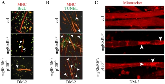
Figure 2. Differentiation of mgRb:Rb−/−:p130−/− DKO myoblasts.
(A) Confocal microscopy analysis for BrdU incorporation in ctrl, mgRb:Rb−/− and mgRb:Rb−/−:p130−/− myotubes at DM-2. Myoblasts were differentiated for 1 day, then exposed to 20 µM BrdU for an additional 16 hr in the presence of growth medium (GM) and immuno-stained for MHC (red) and BrdU (green). Arrowheads label BrdU positive nuclei within myotubes. (B) MHC (red) and TUNEL (green) staining at DM-2. Arrowheads indicate TUNEL positive nuclei, which are invariably located outside myotubes. (C) Mitotracker® (red) staining at DM-2. Arrowheads point to large Mitotracker®-positive perinuclear aggregates.

