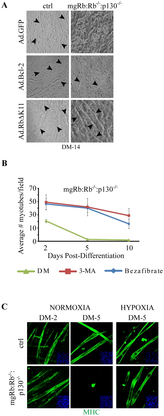
Figure 3. Rescue of mgRb:Rb−/−:p130−/− myogenic defect by autophagy inhibitors and hypoxia.
(A) Brightfield images of mgRb:Rb−/−:p130−/− myoblasts transduced with Ad.GFP, Ad.Bcl-2 or Ad.RbΔK11 and then induced to differentiate for 14 days. Arrowheads point to myotubes. (B) Average number of mgRb:Rb−/−:p130−/− myotubes following treatment with 3-MA, bezafibrate or DM as indicated. Counts are average ± s.d. of 6 fields at 200X (n = 3). (C) Immunostaining for MHC (green) in ctrl and mgRb:Rb−/−:p130−/− cultures differentiated under normoxia or hypoxia. Note myotubes in mgRb:Rb−/−:p130−/− cultures at DM-5 under hypoxia but not normoxia. Nuclei were counterstained with DAPI.

