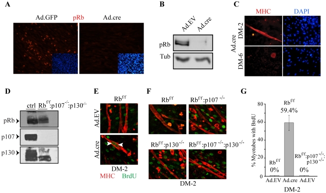
Figure 4. BrdU incorporation analysis of RbΔf versus p107−/−:p130−/− myotubes.
(A) Rbf/f myoblasts were transduced with Ad.GFP or Ad.cre and 48 hr later were immunostained for pRb (red). Nuclei were counterstained with DAPI. (B) Rbf/f myoblasts were transduced with Ad.EV or Ad.cre and immunoblotted for pRb 48 hr later. Tubulin served as loading control. (C) Rbf/f myoblasts were transduced with Ad.cre, induced to differentiate for 2 or 6 days and immunostained for MHC (red). (D) Western blot analysis of pRb, p107 and p130 in skeletal muscle of E16.5 Rbf/f:p107+/−:p130+/− (ctrl) and Rbf/f:p107−/−:p130−/− fetuses. (E) Immunostaining for BrdU and MHC in Ad.EV and Ad.cre transduced Rbf/f myoblasts at DM-2. Myoblasts were differentiated for 1 day, then exposed to 20 µM BrdU for an additional 16 hr in the presence of GM and stained for MHC (red) and BrdU (green). Arrowheads label BrdU positive nuclei within myotubes. (F) Immunostaining for BrdU and MHC in Rbf/f, Rbf/f:p107−/−, Rbf/f:p130−/− and Rbf/f:p107−/−:p130−/− myoblasts at DM-2. Myoblasts were differentiated for 1 day, then exposed to 20 µM BrdU for an additional 16 hr in the presence of GM and stained for MHC (red) and BrdU (green). Note absence of BrdU-positive nuclei in myotubes. (G) Quantification of BrdU incorporation in Ad.EV or Ad.cre transduced Rbf/f and Rbf/f:p107−/−:p130−/− myotubes at DM-2.

