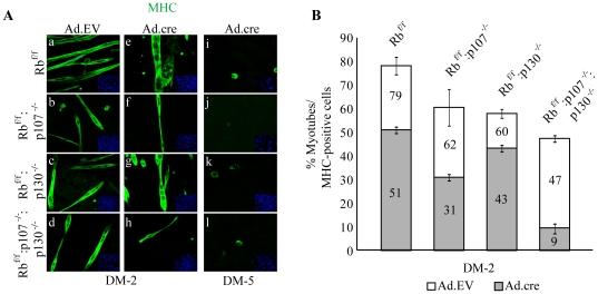
Figure 5. Differentiation potential of double and triple KO myoblasts.
(A) Immunostaining for MHC (green) in Ad.EV and Ad.cre transduced Rbf/f, Rbf/f:p107−/−, Rbf/f:p130−/− and Rbf/f:p107−/−:p130−/− myoblast cultures at DM-2. Nuclei were counterstained with DAPI. (B) Quantification of percent multinucleated myotubes relative to total number of MHC-positive cells (myocytes plus myotubes) in Ad.EV and Ad.cre transduced Rbf/f, Rbf/f:p107−/−, Rbf/f:p130−/− and Rbf/f:p107−/−:p130−/− myoblasts at DM-2 under normoxia. Numbers within bars indicate % for the respective samples.

