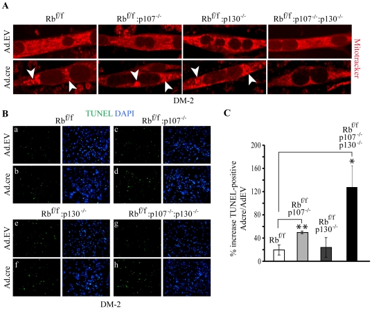
Figure 6. Increased apoptosis associated with differentiation of double and triple KO myoblasts.
(A) Mitotracker® staining of Ad.EV and Ad.cre transduced Rbf/f, Rbf/f:p107−/−, Rbf/f:p130−/− and Rbf/f:p107−/−:p130−/− myoblasts at DM-2. Arrowheads point to large perinuclear aggregates in Ad.cre transduced myotubes. (B) TUNEL staining (green) of Ad.EV and Ad.cre transduced Rbf/f (a–b), Rbf/f:p107−/− (c–d), Rbf/f:p130−/− (e–f) and Rbf/f:p107−/−:p130−/− (g–h) cultures at DM-2. Nuclei were counterstained with DAPI. Note that TUNEL positive nuclei are outside myotubes. (C) Percent increase in TUNEL-positive cells in Ad.cre relative to Ad.EV transduced cultures. Error bars represent s.d. *-p<0.05 and **-p<0.07 t-test comparisons relative to Rbf/f.

