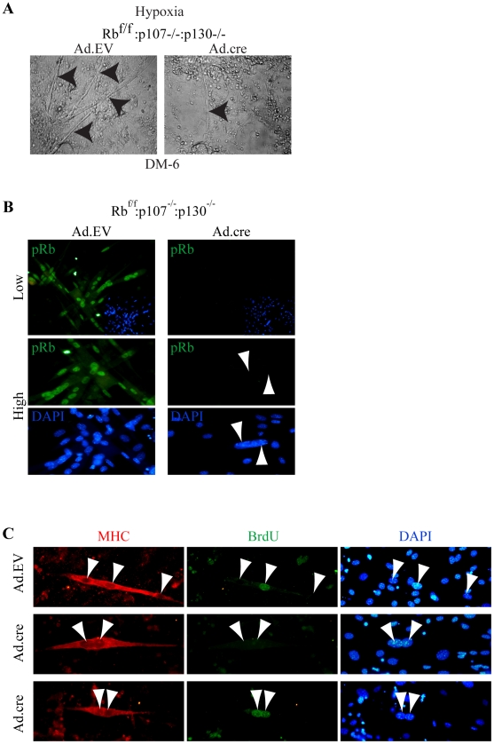
Figure 8. Evidence that bi-nuclear TKO myotubes originate from nuclear duplication, not cell fusion.
(A) Bright-field images of Ad.EV and Ad.cre transduced Rbf/f:p107−/−:p130−/− cultures at DM-6 in hypoxia. Arrowheads point to myotubes. Note the presence of a thin myotube in Ad.cre transduced culture. (B) Top row, low magnification image of Ad.EV or Ad.cre transduced Rbf/f:p107−/−:p130−/− cultures immunostained for pRb (green) at DM-2 (400x). Bottom row, high magnification (630x) of Ad.EV or Ad.cre transduced Rbf/f:p107−/−:p130−/− cultures induced to differentiate and then immunostained for pRb (green), demonstrating absence of detectable pRb within binuclear myocyte. Nuclei were counterstained with DAPI. Arrowheads point to nuclei in the myocyte. (C) Immunostaining for MHC (red) and BrdU (green) at DM-2 of Ad.EV and Ad.cre transduced Rbf/f:p107−/−:p130−/− cultures, which were induced differentiate after equal mixing of BrdU+ labeled and BrdU− myoblast populations. Note mixed BrdU+ and BrdU− nuclei in control myotube (top panel) but only BrdU−:BrdU− or BrdU+:BrdU+ nuclei in TKO myotubes (middle and bottom, respectively).

