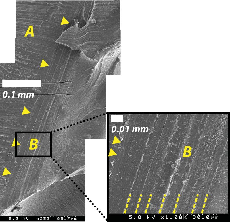
Fig 3.
Scanning electron micrographs of the exposed surface of an initiated crack from case E, oriented approximately vertically as in Fig 1. The external surface of the liner notch (A) features horizontal machining lines. The intersection of the crack with the surface is delineated with triangular markers. The surface of the crack (B) exhibits distinct vertically oriented and parallel clam shell markings (highlighted with dashed lines), verifying the observations made with visible microscopy. The parallel cuts in upper left are artificial marks for locating the fracture surface.

