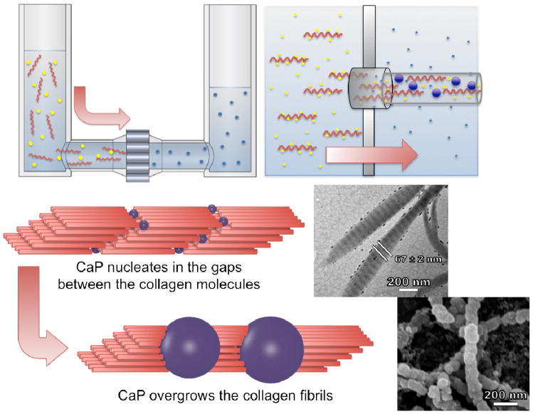
Figure 1.
Experimental setup and proposed model for the formation of mineralized collagen fibrils. Amorphous calcium phosphate formed inside or near the exit of the nanopores simultaneously with the self-assembly of collagen fibrils. The fibrils were extruded from the pores in the direction of the feed solution flow. The upper inset is a transmission electron micrograph of the mineralized collagen fibrils showing visual enhancement of the periodic banding structure as a result of the incorporation of CaP. The lower inset is a scanning electron micrograph of the collagen fibrils showing the overgrowth of CaP.

