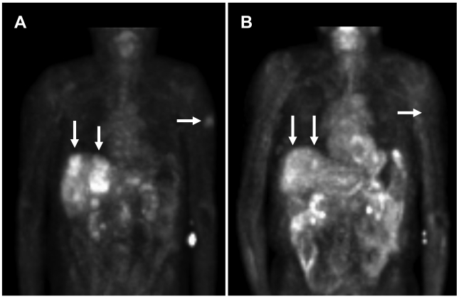
Figure 2.
PET scan demonstrates tumor improvement while on liposomal doxorubicin treatment. A. Patient's PET scan prior to liposomal doxorubicin, and B. Patient's PET scan after 16 cycles (1 cycle = 3 weeks) of liposomal doxorubicin demonstrated shrinkage of tumor with treatment. Note the significant improvement in liver lesions as well as a left upper extremity skin lesion. Arrows indicate tumor lesions with notable reduction with treatment. In the central liver in the region of the caudate lobe there is a lesion which was present on 10/18/05 but no longer seen on 10/23/06 [Standardized Uptake Values (SUV) 10/18/05: Average 6.4, Max 8.3. 10/23/06: Average 2.1, Max 2.6]. In the periphery of the anterior segment of the right lobe there is a lesion which was large on 10/18/05 and much smaller on 10/23/06 [SUV 10/18/05: Average 3.2, Max 4.4. 10/23/06: Average 3.0, Max 3.5].

