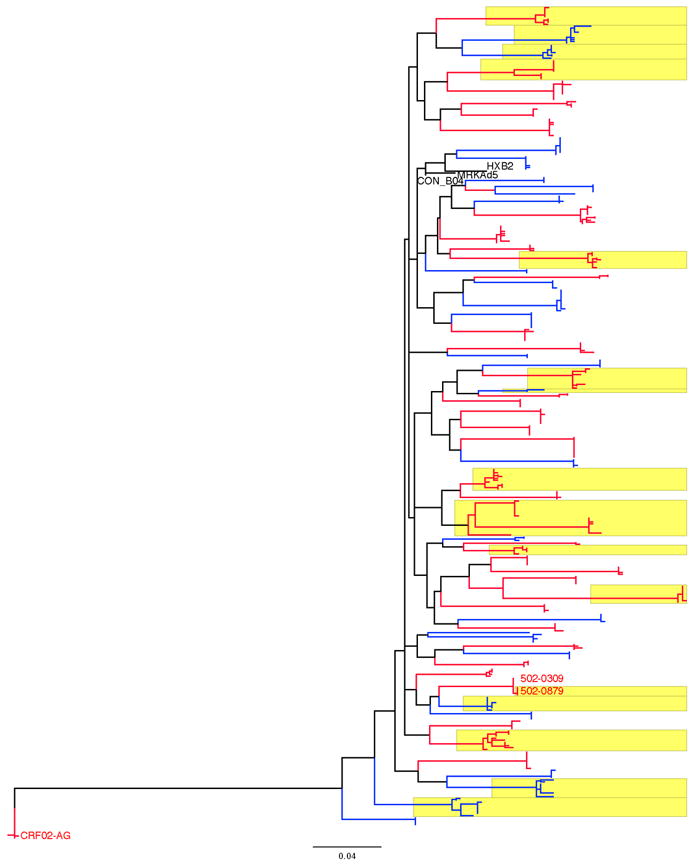
Fig. 1. Maximum-Likelihood phylogenetic tree of gag sequences.

The tree comprises nucleotide sequences from each individual along with the MRKAd5, HXB2 and CON_B04 sequences, and is rooted with sequences from the only subject not infected with a subtype B virus (CRF02-AG). Sequences from placebo recipients are in blue, while sequences from vaccine recipients are in red. Sequences from individuals with two or more founder variants are highlighted in yellow. The sequences from related viruses found in two individuals are labeled with the two subjects’s identification numbers
