
Figure 3. Effect of deleting FGF receptors on the thickness of the lens placode.
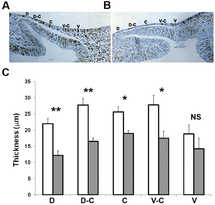
(A, B) Placode thickness was measured at five dorso-ventral locations at E9.5 (mid-placode stage) in wild type (A) and Fgfr1/2CKO (B) placodes. The brown nuclei in these sections are BrdU-labeled. (B) Fgfr1/2CKO placodes (shaded bars) were significantly thinner than wild type placodes (open bars) at most locations. * p<0.05; ** p<0.01; NS, not significantly different