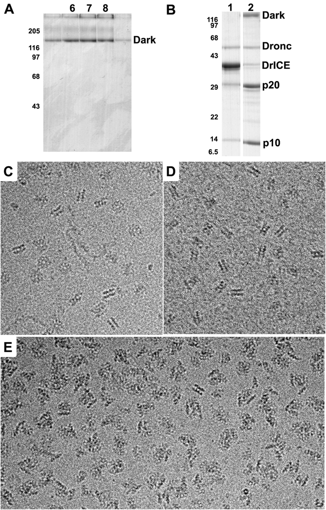
Figure 1. Stability and activity of Dark rings.
A. Glycerol gradient profile of Dark complexes assembled and run in PSB. The peak occurs in fractions 6–8, similar to the migration of the Apaf-1 apoptosome (not shown). Also note that Dark aggregates after heating in SDS gel loading buffer to give a triplet instead of a single band. The positions of molecular weight markers (in kDa) are shown on the left.
B. Proteolysis of DrICE in the absence and presence of Dark complexes. Lane 1: Dronc + DrICE in PSB; Lane 2: co-assembled Dark-Dronc complex + DrICE in PSB; p20 and p10 are cleavage products of DrICE.
C. Frozen-hydrated Dark double-rings at 3 mg/ml in PSB imaged over a hole in the carbon support film.
D. Dark double-rings assembled at 0.5 mg/ml in PSB and imaged on a thin carbon film
E. Dark-Dronc complexes were assembled in PSB and imaged over holes at 3 mg/ml. The images show mostly single-ring aggregates as evidenced by the lack of typical side views for the double-ring and the presence of single-ring edge views.

