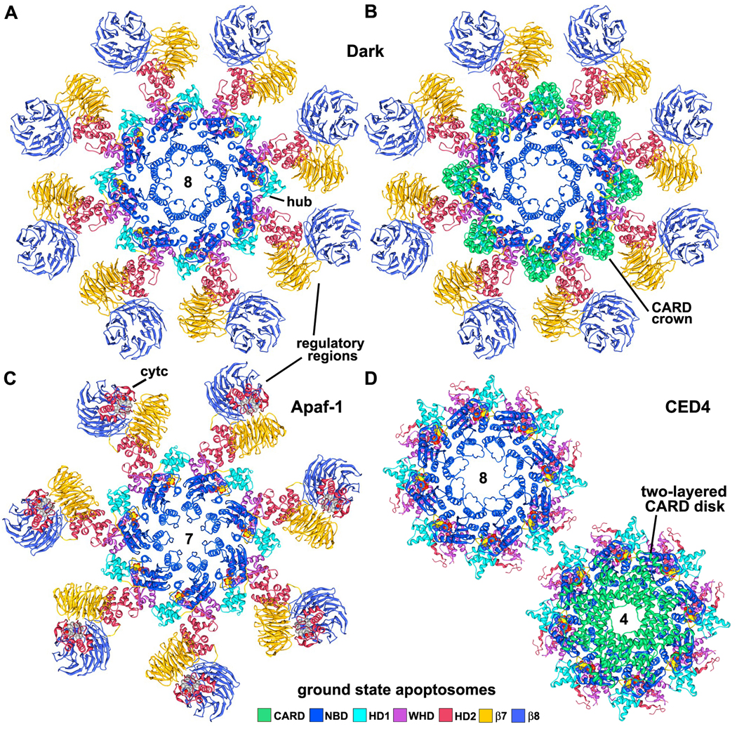
Figure 6. Models of Dark, Apaf-1 and CED-4 apoptosomes.
A. The platform region of the octagonal Dark apoptosome is shown as a molecular ribbon diagram, viewed along the 8-fold axis in a top view.
B. The Dark apoptosome is shown in a top view with the CARDs, to highlight the formation of a CARD crown on the central hub.
C. The heptameric Apaf-1 apoptosome is shown in a top view with bound cytochrome c (although the position of the latter is still being refined; Yuan et al., 2010).
D. (top left) A top view is shown of the octameric CED-4 apoptosome, with CARDs omitted to provide an unobstructed view of the central hub. (bottom right) The CED-4 apoptosome is shown with the double-layered CARD disk, which has 4-fold symmetry (Qi et al., 2010).
See also Figure S8.

