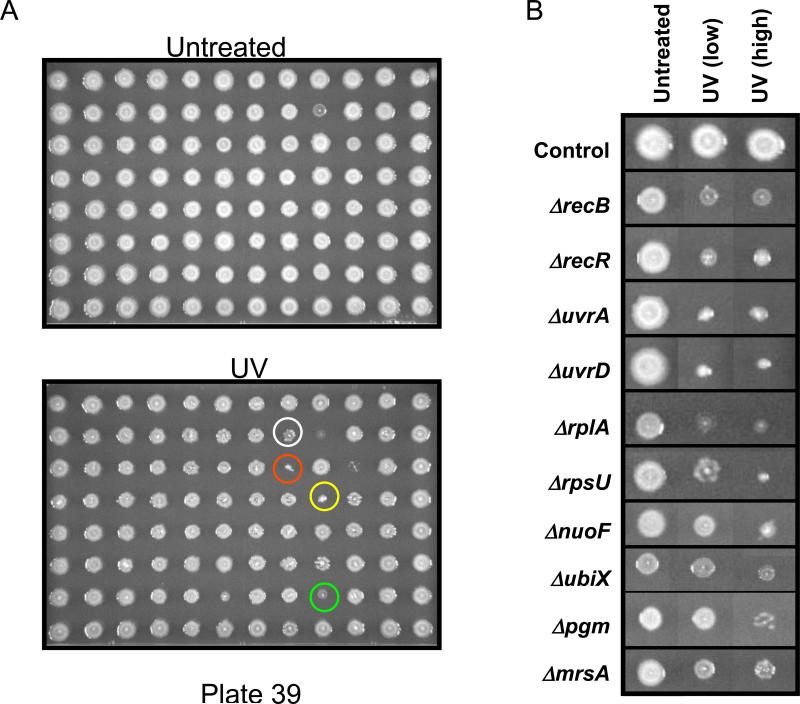
Figure 1. Genomic phenotyping of E. coli mutants with UV.
(A) 96 gene deletion mutants were spotted onto agar plates, left untreated or exposed to two doses of UV, incubated at 37° C for 16 hours and imaged. These doses were used because some cells were only sensitive to the higher dose, while others were sensitive to both conditions. White, red, yellow and green circles identify the UV-sensitive mutants ΔruvA, ΔruvC, ΔuvrA and ΔholC, respectively. (B) Images were taken from many different plates and recompiled to demonstrate that varying degrees of UV-sensitivity were observed in the screen. Examples of a color change from white to grey are shown for ΔrecB and ΔmrsA.

