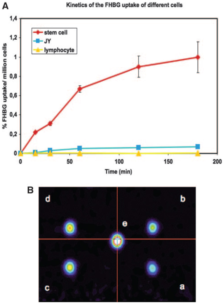
Figure 3.
A, In vitro [18F]-FHBG uptake kinetics based on gamma counting of pig LV-RL-RFP-tTK-MSC in comparison with nontransfected JY human B-lymphoblast and T lymphocytes. Fifteen µCi/mL [18F]-FHBG was incubated with 0.5×10⁶/mL cells for 20 to 180 minutes. Decay corrected activity is expressed as the percentage of the extracellular [18F]-FHBG incorporated per million cells. B, PET of different concentrations (a, 1 ×10⁵ cells/mL; b, 2×10⁵ cells/mL; c, 3×10⁵ cells/ mL; d, 5×10⁵ cells/mL; e, 1×10⁶ cells/mL) of pig LV-RL-RFP-tTK-MSC labeled for 60 minutes.

