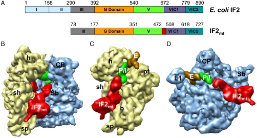
Fig. 1.
Comparison of the domain organization of E. coli IF2 and mammalian IF2mt, and cryo-EM structure of the  complex. (A) Domain alignment of E. coli IF2 and bovine IF2mt. Note that the mature IF2mt (i.e., after deletion of the import sequence) starts from aa residue 78. The 37-aa insertion in IF2mt is highlighted in red. (B) The cryo-EM map of the E. coli 70S ribosome (30S subunit, yellow; 50S subunit, blue) in complex with IF2mt (red), initiator tRNA (green) at the P/I position, and an additional tRNA density (brown, visible in panels C and D) at the ribosomal E site. (C and D) The 30S and 50S subunit portions of the map are shown from their interface sides to reveal the overall binding positions of IF2mt and tRNAs. Landmarks of the 30S subunit: h, head; sh, shoulder; b, body; and pt, platform. Landmarks of the 50S subunit: L1, L1 protein protuberance; CP, central protuberance; and Sb, L7/L12 stalk base. For stereo viewing, see Fig. S2.

