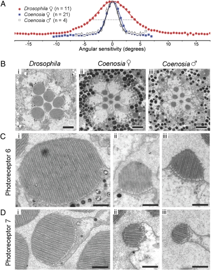
Fig. 3.
Spatial resolution and TEM micrographs of photoreceptors. (A) Spatial resolution of nearly dark-adapted acceptance angle (Δρ) for Drosophila and Coenosia. Mean angular sensitivity functions of female Drosophila and Coenosia ♀ and ♂ data fitted with Gaussians. For Drosophila Δρ = 8.23°, Coenosia ♀ Δρ = 2.88°, and ♂ Δρ = 2.59°. (B) Cross-sections of the distal ommatidia, just below the photoreceptors caps, in the lateral eye regions. (Scale bars, 2 μm.) (C) R6 rhabdomeres are shown as examples for R1–R6 photoreceptors. Male Coenosia rhabdomeres exhibit a “pyramidal” shape. (Scale bars, 500 nm.) See Table S1 for rhabdomere dimensions. (D) R7 Rhabdomeres are shown. (Scale bars, 500 nm.) See Table S1 for rhabdomere dimensions.

