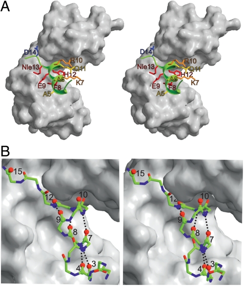
Fig. 1.
Stereo views of the structure of RNase S M13Nle variant consisting of the S-protein (white) and the S-peptide (green) X-ray structure. A shows side chains that were replaced for φ-value analysis. The positions are color coded according to their response to an increase in hydrophobicity with either a strong increase in kon (red), a slight increase in kon (orange), no effect on kon (yellow), or a strong decrease in kon (blue). B shows a ball-and-stick representation of S-peptide bound to S-protein. Oxygens replaced by sulfur for φ-value analysis and the corresponding H-bond donors are marked as balls and the respective helical H-bonds are shown. Coordinates were taken from the X-ray structure (25). The figure was prepared using Molscript (37).

