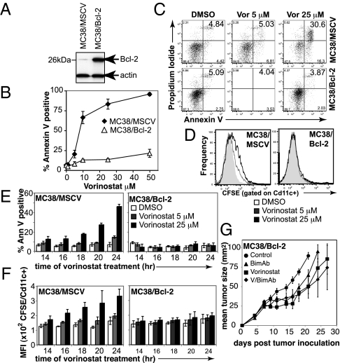
Fig. 2.
Vorinostat-induced apoptosis is required for therapeutic efficacy of V/bimAb. (A) MC38 carcinomas retrovirally transduced to overexpress murine Bcl-2 (MC38/Bcl-2) were assessed for overexpression of exogenous protein by Western blotting. (B) Empty vector control cells (MC38/MSCV) and MC38/Bcl-2 cells were assessed for in vitro sensitivity to vorinostat following culture for 48 h. Data shown are the mean ± SEM of three independent experiments. (C) Tumor cell apoptosis following treatment of MC38/MSCV and MC38/Bcl-2 cells with 5 μM and 25 μM vorinostat for 24 h was assessed using annexinV/propidium iodide staining and flow cytometry. Representative flow cytometry profiles are shown. (D) Engulfment of CFSE-labeled vorinostat-treated (24 h) MC38/MSCV and MC38/Bcl-2 cells by bone marrow-derived CD11c+ APCs was assessed by flow cytometry. Bcl-2 overexpression blocked uptake of MC38 carcinomas following treatment with vorinostat. (E and F) To confirm apoptosis correlated with engulfment by APCs, a time course was performed. Annexin V-positive staining of vorinostat-treated MC38/MSCV and MC38/Bcl-2 cells are shown in E, with the corresponding median fluorescence intensity in FL-1 (i.e., CFSE) following co-culture of tumor cells with APCs, gated on CD11c positive cells shown in F. Data shown are the mean ± SEM of three independent experiments. (G) Efficacy of V/bimAb therapy was then assessed in mice bearing established MC38/Bcl-2 carcinomas. Cohorts of mice with established (>9 mm2) s.c. MC38/Bcl-2 carcinoma were treated with vehicle (n = 6), vorinostat (n = 6), bimAb (n = 6), or V/bimAb (n = 8) using the therapeutic schedule described in Fig. 1. Tumor growth was assessed every 2–3 d; mean tumor size ± SEM are shown. No complete tumor regressions were observed in mice in any of the treatment groups. Data shown are representative of two independent experiments.

