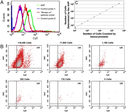
Fig. 4.
Detection of CCRF-CEM cancer cells with the AAP. (A) Flow cytometry assay of CCRF-CEM cells detected by the AAP, in comparison with the results achieved by the “always-on” aptamer probe. The detector voltage used for control probe 1 and the AAP was 540. The detector voltage used for control probe 2 and the always-on probe was 420. (B) Flow cytometry assays of CCRF-CEM cells with decreasing cell amounts in 200 μl binding buffer using the AAP-based cancer cell detection strategy. (C) Calibration curve illustrating the relationship between the amount of CCRF-CEM cells counted by hemocytometer and the number of CCRF-CEM cells detected by flow cytometer with the AAP. Data represent mean ± standard deviation of three cell samples per group.

