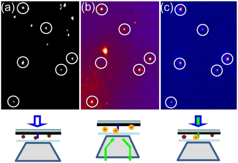
Fig. 3.
Fluorescence imaging of QD-STA using nanopillar illumination. (A) The locations of randomly distributed nanopillars are revealed by bright field imaging. (B) Epi-illumination (through the objective) excites those fluorescent QD-STAs that bind to biotinylated nanopillar surfaces and also those that are nonspecifically stuck on the platinum surface. (C) Fluorescence imaging by nanopillar illumination exclusively excites those QD-STAs that are localized on the nanopillar surfaces with extremely clean fluorescence background. The circles indicate where QD-STA colocalizes with a nanopillar. Schematic illustrations of the microscope setup for three illumination modes are sketched below each image. Note that one of the circled QD-STA is invisible in B due to QD photoblinking.

