Fig. 4.
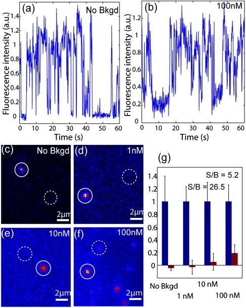
Nanopillar illumination enables single-molecule detection with high fluorescence background. Characteristic single-step photoblinking of QD-STA is evident both at (A) 0 nM and (B) 100 nM of QD-PEG fluorescence background. (C–F) Typical fluorescence images using nanopillar illumination show signal intensity and background intensity at each of the four QD-PEG concentrations 0 nM, 1 nM, 10 nM, and 100 nM. The signal strength is the fluorescence intensity of a QD-STA tethered to the nanopillar in the “on” state (solid circles). The background intensity is the fluorescence intensity at the nanopillar location when the attached QD-STA is in the “off” state (dotted circles). (G) Relative signal (blue) and background (red) intensities at each QD-PEG concentrations.

