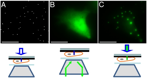
Fig. 6.
Fluorescence imaging using nanopillar illumination in live cells. (A) White-light imaging reveals the locations of nanopillars. (B) Fluorescence imaging by epi-illumination shows the cell shape of a GFP transfected cell. (C) Nanopillar illumination excites only those fluorescence molecules that are very close to nanopillars inside the cell, giving rise to fluorescence spots perfectly colocalized with the nanopillars. Scale bar : 10 μm.

