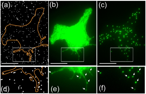
Fig. 7.
Antibody-labeled nanopillars simultaneously recruit and illuminate proteins of interest in live cells. (A) White-light imaging reveals the locations of the nanopillars modified with antibodies against GFP. (B) Fluorescence imaging by epi-illumination shows the shape of a COS7 cell expressing GFP-synaptobrevin. (C) Nanopillar illumination shows extremely clean signal, colocalizing perfectly with the nanopillars inside the cell area. (D–F) Zoom-in images show that nanopillar locations usually have brighter fluorescence compared with surrounding areas, suggesting that some GFP-synaptobrevin proteins were recruited to the nanopillars via GFP and anti-GFP interactions. Scale bar : 10 μm.

