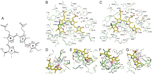
Fig. 1.
Interfacial 1H contacts of the PCB chromophore. (A) Schematic of protein-bound PCB shown in the Pr ZZZssa geometry. The tetrapyrrole rings and representative PCB atoms are labeled for reference. (B and C) Structural views showing residue contacts of the chromophore observed in heteronuclear correlation NMR spectra (see also Figs. S1 and S2) as (B) Pr and (C) Pfr. The presentations for Pr (B) and Pfr (C) states are modeled according to the Cph1 2VEA (21) and PaBphP 3C2W crystal structures (22), respectively. Water locations (W1–W7) are marked as red spheres and numbered (42). (D–G) Close-up views of the 1H contacts of the two D-ring carbons, C19 and C171, as Pr (D and F) and Pfr (E and G). The dashed lines highlight the  interfacial correlations during Pr → Pfr photoconversion. All 1H contacts of the chromophore are summarized in Datasets S3 and S4.

