Fig. 5.
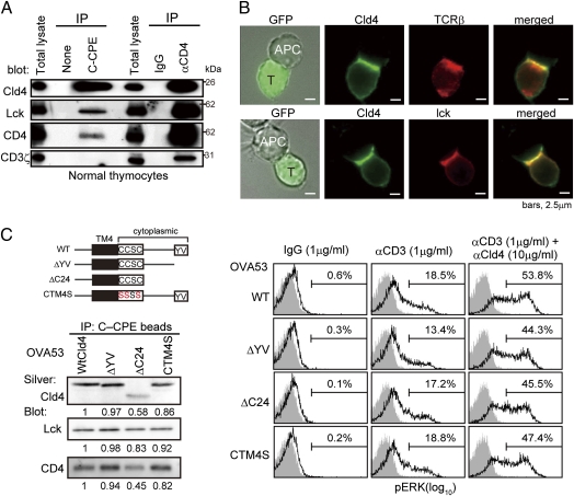
Cld4 is associated with CD4/lck and recruited to immunologic synapse. (A) Newborn B6 thymocytes were lysed and immunoprecipitated with b–C-CPE or b–anti-CD4 and avidin beads followed by immunoblotting with the indicated antibodies. Data are representative of five experiments. (B) OVA53/Cld4 cells were incubated for 6 h with APCs (A20 cells) preloaded overnight with 1 μM OVA, fixed, and immunostained with the indicated antibodies. OVA53/Cld4 cells were identified with GFP. Data are representative of three experiments. (C) OVA53 cells were retrovirally transduced with the mutant Cldn4 as illustrated (Upper Left), and the cell lysates were immunoprecipitated with b–C-CPE and avidin beads followed by immunoblotting with the indicated antibodies. Aliquotes of the immunoprecipitates were electrophoresed and silver-stained (Lower Left). Relative intensities of the signals are indicated. Data are representative of five experiments. OVA53 cells expressing WT or mutant Cld4 were incubated with 1 μg/mL b-hamster IgG, 1 μg/mL b–anti-CD3, or 1 μg/mL b–anti-CD3 plus 10 μg/mL b–anti-Cld4 antibodies followed by cross-linking with 25 μg/mL avidin; 2 min later, ERK phosphorylation was assessed. Data are representative of four experiments (Right).

