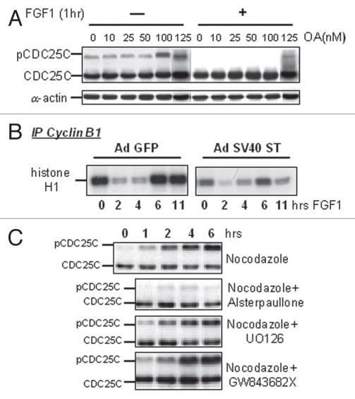
Figure 4.
FGF-induced inhibition of CDC25C phosphorylation is not mediated by activation of a phosphatase. (A) RCS cells were pre-incubated with okadaic acid (OA) at the indicated concentrations for 1 hour and either treated or not with FGF1 for one more hour. 50 µg of total cellular protein were analyzed by SDS-PAGE, followed by immunoblotting for anti-CDC25C antibodies. Equal amount of protein loading was confirmed by α-actin immunodetection. (B) RCS cells were infected with adenoviruses expressing either GFP or SV40 ST antigen following FGF1 treatment as indicated. Kinase activity of immunoprecipitated cyclin B1/CDK1 complexes was assayed in vitro. The cyclin B1/CDK1 complexes were isolated from 0.5 mg of total cellular protein using anti-cyclin B1 antibodies. Histone H1 was used as a substrate. Antimouse IgG was used as a negative control. RCS cells infected with SV40 ST routinely exhibited lower basal levels of cyclin B1-associated kinase activity in untreated cells as assayed in several independent experiments. (C) RCS cells were treated with Nocodazole and either with CDK1 inhibitor (Alsterpaullone), MEK1/2 inhibitor (U0126) or PLK1/3 inhibitor (GW843682X), as marked, and harvested at the indicated times. 50 µg of total cellular protein were analyzed by SDS-PAGE followed by immunoblotting for anti-CDC25C antibodies.

