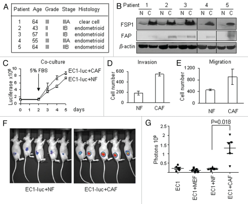
Figure 1.
Characterization of fibroblast cell lines. (A) Clinicopathologic data of the samples used to produce fibroblasts. (B) Western blot analysis of fibroblasts (CAFs and NFs) with anti-Fibroblast Specific Protein 1 (FSP1) and anti-Fibroblast Activation Protein (FAP). N, NF; C, CAF. (C) CAFs stimulate growth of endometrial cancer cell line (EC1) compared to normal fibroblasts in co-culture experiments. (D and E) Conditioned media from CAFs stimulate EC1 cells matrigel invasion (D) and migration (E). Values in (C–E) represent average numbers for five pairs of fibroblasts ± SEM. (F) Images of mice 50 days after injection with EC1-luc cells co-mingled with either NFs or CAFs. (G) Quantification of tumor burden in mice injected with EC1-luc alone or in combination with either mouse embryo fibroblasts (MEFs), endometrial NFs or endometrial CAFs.

