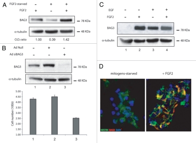
Figure 1.
(A) FGF2 sustains BAG3 protein levels and impacts the proliferation of neural progenitor cells. Western blot analysis of total lysates from adult hippocampus-derived neural progenitor cells (passage number 25) cultured in neurobasal medium (NM) containing N2 supplement plus 20 ng/ml fibroblast growth factor-2 (lane 1), FGF-2-starved cells (lane 2), cells starved and then readministrated with FGF-2 (lane 3). BAG3 endogenous protein was evaluated by imunoblotting with a rabbit polyclonal antibody against BAG3. α-tubulin was detected as normalization control among samples. (B) Adenoviral infection of BAG3 siRNA was performed where indicated at 40 m.o.i. per cell. The cells were FGF2-starved 10 h and then treated with FGF2 for 72 h. Cells were counted and cell lysates were evaluated for BAG3 protein expression. The data reported in (B) are the average of 3 independent experiments. Percentage of viability by trypan blue exclusion did not show significant change among different samples (data not shown). (C) BAG3 expression is induced by EGF and FGF-2 in secondary neurosphere and localizes in nestin posive cells. FGF2 and EGF sustain BAG3 protein expression. Primary neurospheres were prepared from E16 mouse embryos and cultured in neurobasal medium containing N2 supplement plus FGF2 20 ng/ml and EGF 20 ng/ml. After splitting, secondary neurospheres have been cultured for 10 h in N2 supplement (lane 1) or for additional 48 h in N2 + FGF2 (lane 2) or N2 + EGF (lane 3) or N2 + FGF2/EGF (lane 4). (D) Mouse secondary neurospheres where cultured as indicated in (C). Imunofluorescence performed with FITC-labeled anti-nestin antibody and rhodamine-labeled BAG3 antibody are shown in neurobasal medium + N2 cultured cells (left) and neurobasal + N2 + FGF2 20 ng/ml cultured cells (right).

