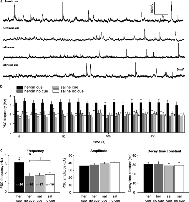
Figure 6.
Cue-induced enhancement of GABAergic inhibition of mPFC pyramidal neurons. (a) Example traces of whole-cell recordings from mPFC pyramidal neurons in control and heroin self-administration animals (n=5 per group) that were re-exposed (cue) or not exposed (no cue) to the cues. Number of neurons in each group: Heroin cue, n=20; Heroin no cue, n=20; Saline cue, n=17; Saline no cue, n=16. (b) Average IPSC frequency in all neurons recorded during a 3-min period (10 s bins). (c) Summary data and statistical analysis: average IPSC frequency was significantly higher in heroin animals on cue re-exposure (*p=0.007). IPCS amplitude and decay time constant remained unaltered (p>0.05) on cue re-exposure. Data represent mean±SEM.

