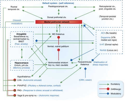
Figure 10.
Anatomical circuits involving the medial prefrontal network (medial prefrontal network) and amygdala. Glutamatergic, presumed excitatory projections are shown in green, GABAergic projections are shown in orange, and modulatory projections in blue. In the model proposed here, dysfunction in the amygdala and/or the medial prefrontal network results in dysregulation of transmission throughout an extended brain circuit that stretches from the cortex to the brainstem, yielding the emotional, cognitive, endocrine, autonomic, and neurochemical manifestations of depression. Intra-amygdaloid connections link the basal and lateral amygdaloid nuclei to the central and medial nuclei of the amygdala and the bed nucleus of the stria terminalis (BNST). Parallel and convergent efferent projections from the amygdala and the medial prefrontal network to the hypothalamus, periaqueductal gray (PAG), nucleus basalis, locus ceruleus, dorsal raphe, and medullary vagal nuclei organize neuroendocrine, autonomic, neurotransmitter and behavioral responses to stressors and emotional stimuli (Davis and Shi, 1999, LeDoux, 2003). In addition, the amygdala and medial prefrontal network interact with the same cortico-striatal-pallidal-thalamic loop, through prominent connections both with the accumbens nucleus and medial caudate, and with the mediodorsal and paraventricular thalamic nuclei, which may function to control and limit responses to stress. Finally, the medial prefrontal network is a central node in the cortical ‘default system' that appears to support self-referential functions such as mood. Other abbreviations: 5-HT—serotonin; ACh—acetylcholine; Cort.—corticosteroid; CRH—corticotrophin releasing hormone; Ctx—cortex; NorAdr—norepinephrine; PVN—paraventricular nucleus of the hypothalamus; PVZ—periventricular zone of hypothalamus; STGr—rostral superior temporal gyrus—VTA—ventral tegmental area.

