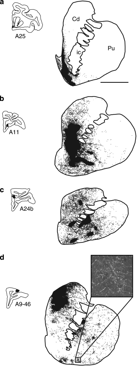
Figure 4.
Schematic chartings of labeled fibers after injections into different prefrontal regions. (a) vmPFC injection site (area 25), (b) OFC injection site (area 11), (c) dACC, (d) dPFC injection site (area 9/46). The focal projection fields are indicated in large solid black shapes. Diffuse projection fibers are found outside of the focal projection fields (as illustrated in the photomicrograph in (d)).

