
Figure 5. CCK-SCAs subthreshold oscillatory preferences.
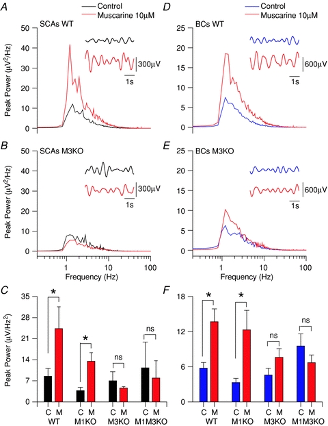
Power spectrum plot of subthreshold activity (holding Vm at −60 mV) for CCK-SCAs WT (A), M3 KO CCK-SCAs (B), WT CCK-BCs (D) and M3 KO CCK-BCs (E) population data in control conditions (black or blue trace) and muscarine 10 μm (red traces). The insets show 5 s holding resting potential raw traces filtered between 0.8 to 2.2 Hz in control (black or blue) and muscarine (red) conditions. C, bar plot of the CCK-SCAs population average data between 1 to 2 Hz for each condition. F, bar plot of the CCK-BCs population average data between 1 to 2 Hz for each condition.